
Library > Medical Assisting > X-Ray > Basic Radiographic Techniques
Try Simtics for free
Start my free trialBasic Radiographic Techniques

Materials Included:
-

-

-

-

-

Check our pricing plans here
Unlimited streaming.
Radiographic technologists assist in the diagnosis and management of human illness by producing diagnostic images (also called radiographs or X-rays) of relevant patient anatomy. This module provides a thorough introduction to basic radiographic techniques, and demonstrates how to set up and perform an X-ray procedure. Including both practice and test modes, the online simulator offers two different patient scenarios, allowing you to learn and practice the steps involved in performing basic radiographic procedures. This module is an ideal resource if you are studying for the American Registry of Radiologic Technologists® (ARRT) registry exams, or if you are just interested in understanding the role of a radiographer.
You’ll learn
- to identify common radiographic equipment and its uses
- the importance of radiation protection for the patient and technologist
- basic radiographic positions, terminology, and positioning methods
- to practice and perfect your skills in preparing for and performing an X-ray procedure
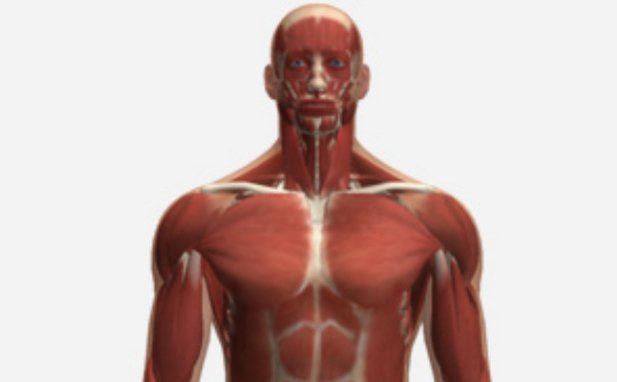
- to better visualize and understand the human anatomy, with our 3D anatomical model
- basic techniques for obtaining optimal images
- techniques for dealing with trauma and pediatric patients
- how to care for the radiographic examining room
- much more (see “content details” for more specific information)
- Discuss and utilize radiographic positioning terminology
- Discuss the care of the radiographic examining room
- Describe common radiographic positions
- Discuss the various approaches to dealing with trauma and pediatric patients
- Discuss basic radiographic positioning methods and steps
- Describe and identify radiographic equipment
- Describe positioning landmarks
- Describe and explain the significance of obtaining a pertinent patient history
- Describe and explain the technologists' role with respect to patient safety and ALARA
- Discuss the importance of radiation protection
- Discuss infection control and prevention
- Describe and explain the reason for patient breathing techniques in order to obtain optimum radiographic images
- Analyze the radiographs for quality and proper positioning criteria
- Explain the Patient's Bill of Rights, HIPAA Privacy Rule (HIPAA), and Patient Safety Act (see reference)
The SIMTICS modules are all easy to use and web-based. This means they are available at any time as long as the learner has an internet connection. No special hardware or other equipment is required, other than a computer mouse for use in the simulations. Each of the SIMTICS modules covers one specific procedure or topic in detail. Each module contains:
- an online simulation (available in Learn and Test modes)
- descriptive text, which explains exactly how to perform that particular procedure including key terms and hyperlinks to references
- 2D images and a 3D model of applied anatomy for that particular topic
- a step by step video demonstration by an expert
- a quiz
- a personal logbook that keeps track of all the modules the learner has studied and how long
For more details on features and how your students can benefit from our unique system, click here.


