
Library > Medical Professional - Ultrasound > Cardiac > Basic Echocardiography Views for Medical Professionals
Try Simtics for free
Start my free trialBasic Echocardiography Views for Medical Professionals

Check our pricing plans here
Unlimited streaming.
This module follows on from Basic Echocardiography Techniques and teaches you how to obtain the views required to examine all the anatomy of the heart. Including both Learn and Test modes, the online simulator offers three clinical scenarios that test your ability to obtain basic echocardiography views of the heart. Practice the steps of the procedures online as often as you want, until you feel confident.
If you are not a medical student or physician, you may prefer the other version of this module, which includes all the procedural information needed by professionals in other roles: /shop/imaging/sonography/echocardiography/basic-echocardiography-views
You’ll learn
- the use of scanning planes and transducer positions to obtain optimal images
- appropriate patient positioning and breathing techniques for cardiac ultrasound examinations
- to identify and obtain images of the heart, ascending aorta, main pulmonary arteries, and great veins
- to practice, perfect, and test your skills in performing an echocardiographic assessment of the heart
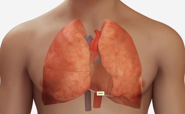
- to better visualize and understand the anatomy and physiology of the heart and major vessels, with our 3D model and illustrations
- to differentiate normal and abnormal sonographic appearances of the heart
- to obtain measurements of various structures within the heart
- much more (see Content Details for more specific information)
- Describe and explain heart anatomy
- Describe and explain heart physiology
- List indications for ultrasound of the heart
- Echocardiography protocol:
- Describe and explain the reason for patient preparation for echocardiography
- Describe and explain transducer and preset selection
- Describe and demonstrate the reason for patient positioning for echocardiography
- Describe and demonstrate the use of scanning planes and the echocardiography protocol
- Demonstrate different transducer positions to obtain optimal images
- Describe and explain the reason for patient breathing techniques in order to obtain optimum images
- Identify and obtain images of heart, ascending aorta, main pulmonary arteries, and great veins
- Obtain measurements of various structures within the heart
- Differentiate normal and abnormal sonographic appearances of the heart
- Explain the Patient's Bill of Rights, HIPAA Privacy Rule, and Patient Safety Act (see reference)
The SIMTICS modules are all easy to use and web-based. This means they are available at any time as long as the learner has an internet connection. No special hardware or other equipment is required, other than a computer mouse for use in the simulations. Each of the SIMTICS modules covers one specific procedure or topic in detail. Each module contains:
- an online simulation (available in Learn and Test modes)
- descriptive text, which explains exactly how to perform that particular procedure including key terms and hyperlinks to references
- 2D images and a 3D model of applied anatomy for that particular topic
- a step by step video demonstration by an expert
- a quiz
- a personal logbook that keeps track of all the modules the learner has studied and how long
For more details on features and how your students can benefit from our unique system, click here.


