
Library > Medical Professional - Ultrasound > Cardiac > Doppler Techniques for Medical Professionals
Try Simtics for free
Start my free trialDoppler Techniques for Medical Professionals

Check our pricing plans here
Unlimited streaming.
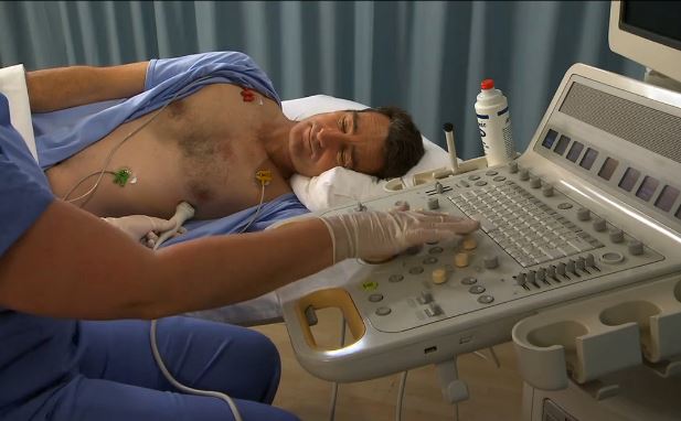
Doppler techniques are used to monitor blood flow through the heart and major vessels. This module teaches you how to prepare for and perform spectral and color Doppler scans of the heart. Including both Learn and Test modes, the online simulator offers three clinical scenarios that cover different situations you might encounter in the clinical environment and tests your ability to learn Doppler techniques. Practice the steps of the procedures online as often as you want, until you feel confident.
If you are not a medical student or physician, you may prefer the other version of this module, which includes all the procedural information needed by professionals in other roles: /shop/imaging/sonography/echocardiography/doppler-techniques-and-instrumentation/
You’ll learn
- to practice, perfect, and test your skills in the use of spectral Doppler and color Doppler instrumentation
- to identify and describe anatomy represented on spectral Doppler tracings
- to use tissue Doppler and identify tissue Doppler wave forms
- to better visualize and understand the anatomy and physiology of the heart and major vessels, with our 3D model and illustrations
- to differentiate on color images the normal and abnormal sonographic appearances of the heart
- to obtain measurements of various structures within the heart
- much more (see Content Details for more specific information).
- Review normal cardiac anatomy.
- Describe and demonstrate the use of spectral Doppler and color Doppler instrumentation.
- Identify and describe anatomy represented on spectral Doppler tracings.
- Differentiate between normal and abnormal anatomy represented on color images.
- Describe and demonstrate tissue Doppler.
- Describe and identify tissue Doppler wave forms.
- Define and use related medical terminology.
- Explain the Patient Privacy Rule (HIPAA) and Patient Safety Act.
The SIMTICS modules are all easy to use and web-based. This means they are available at any time as long as the learner has an internet connection. No special hardware or other equipment is required, other than a computer mouse for use in the simulations. Each of the SIMTICS modules covers one specific procedure or topic in detail. Each module contains:
- an online simulation (available in Learn and Test modes)
- descriptive text, which explains exactly how to perform that particular procedure including key terms and hyperlinks to references
- 2D images and a 3D model of applied anatomy for that particular topic
- a step by step video demonstration by an expert
- a quiz
- a personal logbook that keeps track of all the modules the learner has studied and how long
For more details on features and how your students can benefit from our unique system, click here.


