
Library > Medical Professional - Ultrasound > OB/GYN > Ultrasound Assessment of Fetal Growth and High-Risk Pregnancies for Medical Professionals
Try Simtics for free
Start my free trialUltrasound Assessment of Fetal Growth and High-Risk Pregnancies for Medical Professionals

Check our pricing plans here
Unlimited streaming.
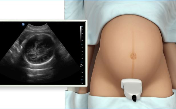
Obstetric ultrasound is the most powerful way to assess the fetus. This module teaches you how to prepare for and perform an ultrasound assessment of fetal growth and high-risk pregnancies. Including both Learn and Test modes, the online simulator offers three clinical scenarios that you might encounter in a clinical situation. Practice the steps of the procedures online as often as you want, until you feel confident.
If you are not a medical student or physician, you may prefer the other version of this module, which includes all the procedural information needed by professionals in other roles: /shop/imaging/sonography/obstetrics/ultrasound-assessment-of-fetal-growth/
You’ll learn
- to practice, perfect, and test your skills in performing an ultrasound scan to assess fetal growth and high-risk pregnancies
- to better visualize and understand the key structures and components of the fetus at various stages of development during the second and third trimesters, with our 3D model and illustrations
- the criteria and testing of fetal well-being, including the biophysical profile
- how to evaluate normal and abnormal findings relating to the amniotic fluid, placenta, and umbilical cord, and abnormalities that may occur with multiple pregnancies
- how intrauterine growth restriction is evaluated by ultrasound
- about peri- and post-pregnancy maternal diseases and complications and how to compare immune and non-immune hydrops
- much more (see Content Details for more specific information)
- Describe and demonstrate how to evaluate normal and abnormal findings relating to the amniotic fluid, placenta, and umbilical cord.
- Describe the use of Doppler in evaluating fetoplacental circulation.
- Give the criteria and demonstrate the testing of fetal well-being, including the biophysical profile.
- Describe techniques for the sonographic evaluation of high-risk pregnancies including multiple gestations.
- List and describe peri- and post-pregnancy maternal diseases and complications and be able to compare immune and non immune hydrops.
- Explain how intrauterine growth restriction is evaluated by ultrasound.
- Describe and recognize on images abnormalities that may occur with multiple pregnancies.
- Define and use related medical terminology.
The SIMTICS modules are all easy to use and web-based. This means they are available at any time as long as the learner has an internet connection. No special hardware or other equipment is required, other than a computer mouse for use in the simulations. Each of the SIMTICS modules covers one specific procedure or topic in detail. Each module contains:
- an online simulation (available in Learn and Test modes)
- descriptive text, which explains exactly how to perform that particular procedure including key terms and hyperlinks to references
- 2D images and a 3D model of applied anatomy for that particular topic
- a step by step video demonstration by an expert
- a quiz
- a personal logbook that keeps track of all the modules the learner has studied and how long
For more details on features and how your students can benefit from our unique system, click here.


