
Library > Radiography > Radiography Theory > Computed Tomography and Magnetic Resonance Imaging
Try Simtics for free
Start my free trialComputed Tomography and Magnetic Resonance Imaging

Materials Included:
-

-

-

-

Check our pricing plans here
Unlimited streaming.
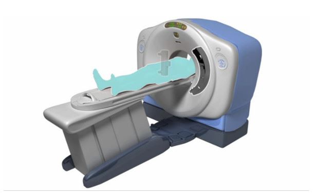
Computed tomography (CT) and magnetic resonance imaging (MRI) are imaging technologies that create cross sections or "slices" of the body part being scanned. This module covers the basic principles of CT, spiral CT and MRI scanning, the differences between them, and advantages of each. It provides an understanding of the technology and techniques involved in using these advanced imaging modalities. If you are studying for the American Registry of Radiologic Technologists® (ARRT) registry exams, this module is ideal.
You’ll learn
- what computed tomography is and how it is achieved
- the differences between conventional tomography, computed tomography, and spiral computed tomography
- to identify the different components of a CT system and visualize how a CT system works
- to describe how MRI works, and its indications and contraindications
- much more (see “content details” for more specific information)
- Explain the purpose, principles, and application of computed tomography (CT)
- Discuss the concepts of transverse tomography, translation, and reconstruction of images
- Discuss the design features that make spiral computed tomography possible
- Explain the spiral imaging principles of interpolation, pitch, index, and section sensitivity
- Identify the appearance of contrast media on a CT image
- Identify basic cross-sectional anatomy of the head, thorax, and abdomen on diagrams, magnetic resonance imaging (MRI), and CT images
- Explain the purpose, principles, and application of MRI
- Identify the appearance of T1 and T2 MRI images
The SIMTICS modules are all easy to use and web-based. This means they are available at any time as long as the learner has an internet connection. No special hardware or other equipment is required, other than a computer mouse for use in the simulations. Each of the SIMTICS modules covers one specific procedure or topic in detail. Each module contains:
- an online simulation (available in Learn and Test modes)
- descriptive text, which explains exactly how to perform that particular procedure including key terms and hyperlinks to references
- 2D images and a 3D model of applied anatomy for that particular topic
- a step by step video demonstration by an expert
- a quiz
- a personal logbook that keeps track of all the modules the learner has studied and how long
For more details on features and how your students can benefit from our unique system, click here.


