US Library > Sonography > Echocardiography > Electrocardiogram (ECG) 12-Lead

Try Simtics for free

Start my free trial

Electrocardiogram (ECG) 12-Lead

Sonography > Echocardiography
Ecg12

Materials Included:

  • Ecg12 Text
  • Ecg12 Video
  • Ecg12 Anatomy
  • Ecg12 Simulation
  • Ecg12 Quiz
Learn how to prepare for, record and interpret a 12-lead ECG.

Check our pricing plans here

An electrocardiogram (ECG or EKG) is a method to graphically display the electric current generated by the heart muscle during a heartbeat. This SIMTICS module teaches you how to prepare for, record and interpret a 12-lead ECG.  The interactive simulator lets you learn and practice how to record an electrocardiogram, and how to interpret the heart rate and rhythms for common pathologies. Online simulations offer a safe practice environment and also enable you to try out procedures that you might not otherwise experience. This module is ideal if you are studying for the American Registry for Diagnostic Medical Sonography (ARDMS) registry exams.

You’ll learn

  • to practice, perfect and test your skills in setting up, recording and interpreting a 12-lead ECG
  • to better visualize and understand the anatomy and physiology of the heart, with our 3D model and illustrations
  • how to determine the heart rate and identify different heart rhythms on the ECG strip: atrial, junctional, ventricular, and pacemaker rhythms, and AV blocks
  • about stress testing and Holter monitoring
  • much more (see Content Details for specific information)
Introduction
Step 1 - Preparation
Step 1.1 - Tray preparation
Step 1.2 - Patient preparation
Step 1.3 - Operator preparation
Step 2 - Place electrodes
Step 2.1 - Place the limb electrodes
Step 2.1.1 - Bipolar limb leads
Step 2.1.2 - Unipolar limb leads: augmented leads
Step 2.2 - Place the chest electrodes
Step 2.2.1 - Unipolar leads: Chest leads
Step 3 - Record the ECG
Step 3.1 - Enter the patient's details into the machine
Step 3.2 - Check the machine is calibrated
Step 3.3 - Record the ECG
Step 3.4 - Annotate the presence of symptoms on the ECG tracing
Step 4 - Analyze the rhythm strip (2 lead)
Step 4.1 - Assess the rate
Step 4.1.1 - The 6-second ECG count
Step 4.1.2 - Count large squares
Step 4.1.3 - Count small squares
Step 4.1.4 - Sequence method
Step 4.2 - Assess the rhythm
Step 4.2.1 - Ventricular rhythm
Step 4.2.2 - Atrial rhythm
Step 4.2.3 - Regularity
Step 4.3 - Identify and assess the P wave
Step 4.4 - Assess the intervals (conduction)
Step 4.4.1 - PR interval
Step 4.4.2 - QRS duration
Step 4.4.3 - QT interval
Step 4.5 - Evaluate overall appearance
Step 5 - Sinus rhythms
Step 5.1 - Features of sinus rhythms
Step 5.2 - Sinus bradycardia
Step 5.3 - Sinus tachycardia
Step 5.4 - Sinus arrhythmia
Step 5.5 - Sinoatrial block
Step 5.6 - Sinus arrest
Step 6 - Atrial arrhythmia
Step 6.1 - Premature atrial complexes (PACs)
Step 6.2 - Wandering atrial pacemaker
Step 6.3 - Multifocal atrial tachycardia
Step 6.4 - Supraventricular tachycardia
Step 6.4.1 - Atrial tachycardia
Step 6.4.2 - AVNRT
Step 6.4.3 - AVRT
Step 6.5 - Atrial flutter
Step 6.6 - Atrial fibrillation
Step 7 - Junctional arrhythmia
Step 7.1 - Premature junctional complexes (PJCs)
Step 7.2 - Junctional escape beats/rhythm
Step 7.3 - Accelerated junctional rhythm
Step 7.4 - Junctional tachycardia
Step 8 - Ventricular arrhythmia
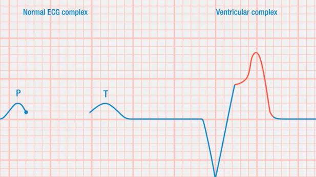
Step 8.1 - Premature ventricular complexes
Step 8.1.1 - Types of PVC
Step 8.2 - Ventricular escape beats
Step 8.3 - Idioventricular rhythm
Step 8.4 - Accelerated idioventricular rhythm (AIVR)
Step 8.5 - Ventricular tachycardia (VT)
Step 8.5.1 - Types of VT
Step 8.6 - Ventricular fibrillation (VF)
Step 8.7 - Asystole
Step 8.8 - Pulseless electrical activity
Step 9 - AV blocks
Step 9.1 - First-degree AV block
Step 9.2 - Second-degree AV block
Step 9.2.1 - Second-degree AV block type I (Wenckebach, or Mobitz type I)
Step 9.2.2 - Second-degree AV block type II (Mobitz type II)
Step 9.2.3 - Second-degree AV block, 2:1 conduction (2:1 AV block)
Step 9.3 - Third-degree/complete AV block
Step 10 - Pacemaker rhythms
Step 10.1 - Pacemaker terminology
Step 10.2 - Pacemaker systems
Step 10.2.1 - Single-chamber pacemakers
Step 10.2.2 - Dual-chamber pacemakers
Step 10.2.3 - Transcutaneous pacing
Step 10.3 - Pacemaker malfunction and complications
Step 10.4 - Analyzing pacemaker function with an ECG
Step 11 - Interpret the 12-lead ECG
Step 11.1 - Normal 12-lead ECG
Step 11.1.1 - Leads
Step 11.1.2 - Layout of the 12-lead ECG
Step 11.2 - Axis
Step 11.2.1 - Vectors
Step 11.2.2 - Einthoven's triangle/Hexaxial reference system
Step 11.2.3 - Two-lead method of axis determination
Step 11.3 - Myocardial ischemia
Step 11.3.1 - ST segment changes
Step 11.3.2 - T wave changes
Step 11.4 - Myocardial infarction
Step 11.4.1 - ST changes
Step 11.4.2 - T wave changes
Step 11.4.3 - Q waves
Step 11.5 - Pericarditis
Step 11.6 - Pericardial effusion
Step 11.7 - Electrolyte imbalance
Step 11.7.1 - Imbalance of sodium ions
Step 11.7.2 - Imbalance of calcium ions
Step 11.7.3 - Imbalance of magnesium ions
Step 11.7.4 - Imbalance of potassium ions
Step 11.8 - Conduction abnormalities
Step 11.8.1 - Right bundle branch block
Step 11.8.2 - Left bundle branch block
Step 11.9 - Analyzing an ECG
Step 11.9.1 - Systematic method
Step 12 - Stress testing and Holter monitoring
  • Describe the anatomy and physiology of the heart
  • Identify the basic electrophysiology of the heart
  • Describe the sinus mechanisms
  • Describe atrial rhythms
  • Describe junctional rhythms
  • Describe ventricular rhythms
  • Describe atrioventricular (AV) blocks
  • Describe pacemaker rhythms
  • Explain and demonstrate the 12-lead ECG
  • Describe stress testing and Holter monitoring

The SIMTICS modules are all easy to use and web-based. This means they are available at any time as long as the learner has an internet connection. No special hardware or other equipment is required, other than a computer mouse for use in the simulations. Each of the SIMTICS modules covers one specific procedure or topic in detail. Each module contains:

  • an online simulation (available in Learn and Test modes)
  • descriptive text, which explains exactly how to perform that particular procedure including key terms and hyperlinks to references
  • 2D images and a 3D model of applied anatomy for that particular topic
  • a step by step video demonstration by an expert
  • a quiz
  • a personal logbook that keeps track of all the modules the learner has studied and how long

For more details on features and how your students can benefit from our unique system, click here.

Don't wait. Try SIMTICS now!

Try Simtics for free

Start my free trial