
Library > Radiography > Radiography Theory > Computer Science, the Digital Image, and Computed Radiography
Try Simtics for free
Start my free trialComputer Science, the Digital Image, and Computed Radiography

Materials Included:
-

-

-

Check our pricing plans here
Unlimited streaming.
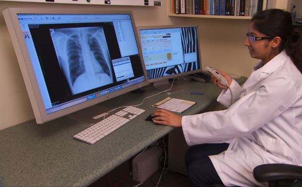
Medical imaging is dependent on the computer for most aspects of acquiring and processing the image, and for subsequent transmission and storage of the recorded data. This theory module provides an introduction to computer science, the technology used in radiographic imaging, and the difference between digital imaging and computed radiography. It also covers the elements of the digital image and how these affect image quality. If you are studying for the American Registry of Radiologic Technologists® (ARRT) registry exams, this module is an ideal learning resource.
You’ll learn
- to identify the different components of a medical imaging computer system
- to describe the fundamentals of digital imaging, and how digital imaging compares with film screen imaging
- to identify the different elements of a digital image and their effect on image quality
- how to explain the science behind digital and computed radiography
- much more (see “content details” for more specific information)
- Review history and development of the computer.
- Define terminology associated with digital imaging systems.
- Describe the various types of digital receptors.
- Discuss the fundamentals of digital radiography, distinguishing between cassette-based systems and cassette-less systems.
- Describe the advantages and disadvantages of digital imaging versus cassette-based imaging.
- Discuss spatial resolution, contrast resolution, and noise related to digital imaging.
- Describe the features of a contrast-detail curve, and interpret a modulation transfer function curve.
- Discuss how post-processing allows the visualization of a wide dynamic range.
- Explain the relevant features of a storage phosphor imaging plate and operating characteristics of a computed radiography reader.
- Describe how digital imaging can be used to help reduce patient radiation dose.
- Describe HIPAA concerns with electronic information.
- Explain the Patient's Bill of Rights, Patient Privacy Rule (HIPAA) and Patient Safety Act.
The SIMTICS modules are all easy to use and web-based. This means they are available at any time as long as the learner has an internet connection. No special hardware or other equipment is required, other than a computer mouse for use in the simulations. Each of the SIMTICS modules covers one specific procedure or topic in detail. Each module contains:
- an online simulation (available in Learn and Test modes)
- descriptive text, which explains exactly how to perform that particular procedure including key terms and hyperlinks to references
- 2D images and a 3D model of applied anatomy for that particular topic
- a step by step video demonstration by an expert
- a quiz
- a personal logbook that keeps track of all the modules the learner has studied and how long
For more details on features and how your students can benefit from our unique system, click here.


